
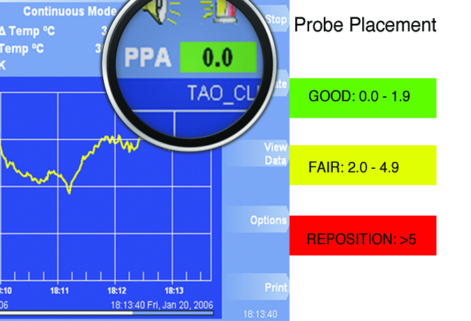
- Operates in conjunction with the Bowman Perfusion Monitor®
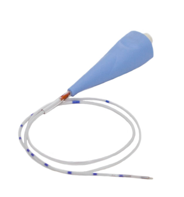
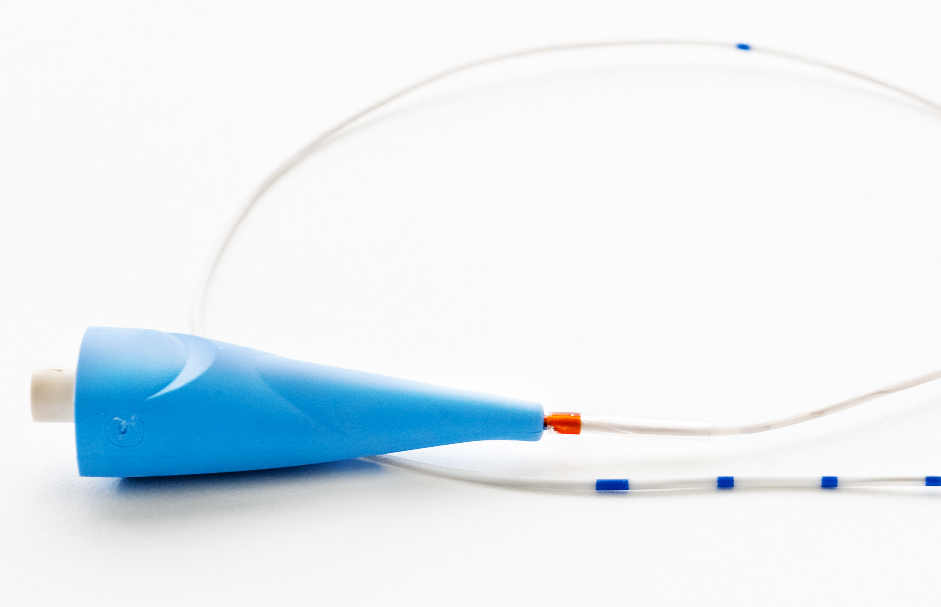
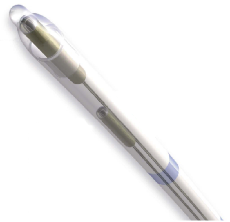
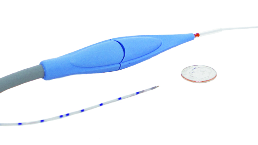
- Biocompatible, flexible catheter with a ~1 mm (3 French/19 gauge) diameter
- CT compatible
- Centimeter markings for verification of depth.
- Color-coded to facilitate connection with the umbilical cord
- Compatible with the QFlow 500™ Titanium Bolts
- No zeroing or calibration required
- FDA cleared for 10 days use in situ
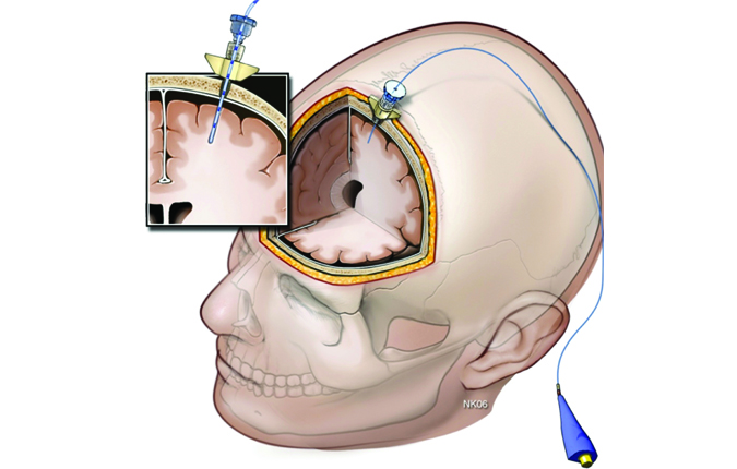
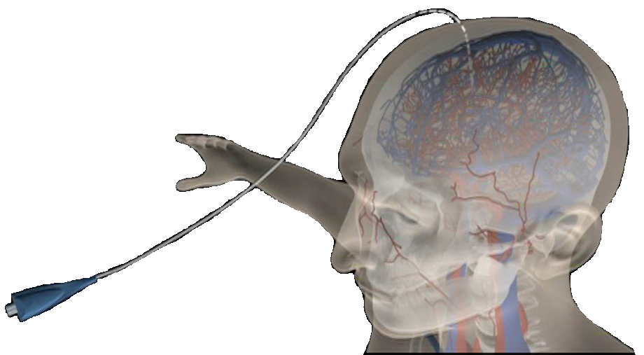
How do you insert the Perfusion Probe?
Similar to other brain parenchymal probes, the QFlow 500™ Perfusion Probe is inserted through a burr hole and positioned 2.5-3cm below the dura into white matter. For optimal fixation Hemedex recommends using the QFlow 500™ Titanium Bolts.
Where do you insert the Perfusion Probe?
The probe should be placed in the vascular territory of interest:
- In areas which are at risk for ischemia.
- In watershed area of vessels likely to experience vasospasm.
- In any area in which you are interested in measuring Cerebral Blood Flow (CBF).
The probe tip should be completely surrounded by tissue.
Is the Perfusion Probe reusable?
No, the QFlow 500™ Perfusion Probe is a single patient use item.
What is the maximum time a Perfusion Probe can be left in situ?
The probe is indicated for 10 days of use.
Is the Perfusion Probe CT compatible?
Yes.
Is the Perfusion Probe MRI compatible?
No.
Does the local measurement reflect the regional blood flow?
It has been demonstrated in one study that the local perfusion measurements reflect regional changes of flow.
“In this report, we demonstrate that (Thermal Diffusion Flowmetry)TDF allows the assessment of cerebral hemodynamic parameters, such as perfusion and vascular resistance, in patients with SAH and reliably detects the development of vasospasm-associated hypoperfusion. Furthermore, we determine the diagnostic cutoff values, predictive values, and likelihood ratios of TDF for identification of symptomatic vasospasm. Finally, we analyze whether multifocal rCBF monitoring increases diagnostic sensitivity in the identification of symptomatic vasospasm when compared to the use of a single probe.” “Regional cerebral blood flow monitoring in the diagnosis of delayed ischemia following aneurysmal subarachnoid hemorrhage” Peter Vajkoczy, M.D., Peter Horn, M.D., Claudius Thomé, M.D., Elke Munch, M.D., and Peter Schmiedek, M.D.J. Neurosurg./Volume98/June 2003
However, this probe takes a local (approximately 0.3 ml of tissue) measurement of blood flow, and regional flow extrapolation from the local blood flow measurement is not claimed.
How does the probe measure perfusion?
The probe uses a thermal diffusion technique. For more information click here.